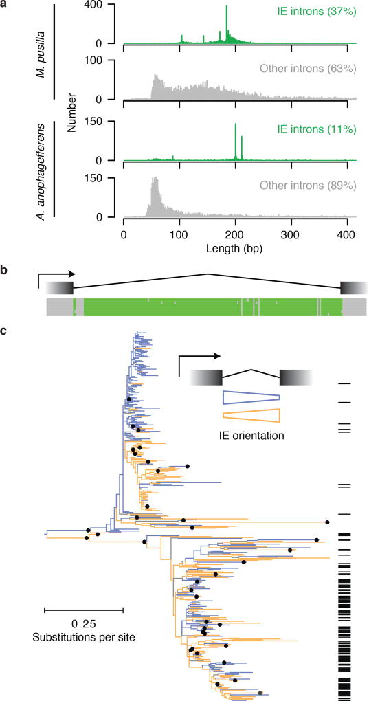
Figure 2. Identification of IEs in A. anophagefferens.
a, Validated lengths for IE (blue) and other (gray) introns. b, A. anophagefferens IEs share sequence similarity in intronic, not in neighboring exonic sequence. Six example IEs contain regions with maximal pairwise identities from 96 to 100%. Bases position identities in at least 5 of the 6 sequences are green. c, Most A. anophagefferens IEs can be aligned to form one or more related groups. Nodes present in >50% of 1,000 bootstraps are indicated with black dots on the ML tree. IEs are found in either orientation with respect to the intron (orange and blue). Many elements carry 3′ splice sites in both orientations (black lines at right).

