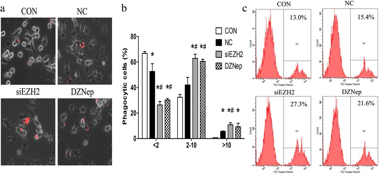
Fig. 4.

Downregulation of EZH2 in GBM enhances microglia phagocytosis. a Murine GL261 cells were pre-treaded with siEZH2 and DZNep for 24 h and then co-cultured with primary microglia (PM) for another 24 h. The fluorescent latex beads were added to microglia for 90 min. Then, the phagocytosis of microglia, indicated as red fluorescence, was observed by confocal microscope. Representative pictures of phagocytic microglia were illustrated; scale bar = 20 μm. b Quantification of PM amounts with phagocytic activity as shown in a. Microglia were divided into three groups in virtue of fluorescent beads contained within PM (< 2 beads/cell, 2–10 beads/cell, and > 10 beads/cell) in 20 randomly selected fields. Data are presented as the mean ± SD from three experiments performed on independently derived microglia cultures. c Murine GL261 cells were pre-treaded with siEZH2 and DZNep for 24 h and then co-cultured with primary microglia for another 24 h. The fluorescent latex beads were added to microglia for 90 min. Amounts of latex beads phagocytized by microglia were detected by flow cytometry. NC and PBS were set as controls, respectively, and nothing were added to Con group. All these experiments were repeated thrice. *p < 0.05 vs. MG group; #p < 0.05 vs. GCM group
