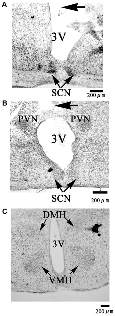
Fig. 5.
Photomicrographs of representative coronal brain sections including the SCN (A, B), PVN (B), DMH and VMH (C) of a mouse received ICV injection. The injection tract (arrow) was described in the panel (A, B). 3V, third ventricle; SCN, suprachiasmatic nucleus; PVN, paraventricular nucleus; DMH, dorsomedial hypothalamus; VMH, ventromedial hypothalamus.

