Figure 2.
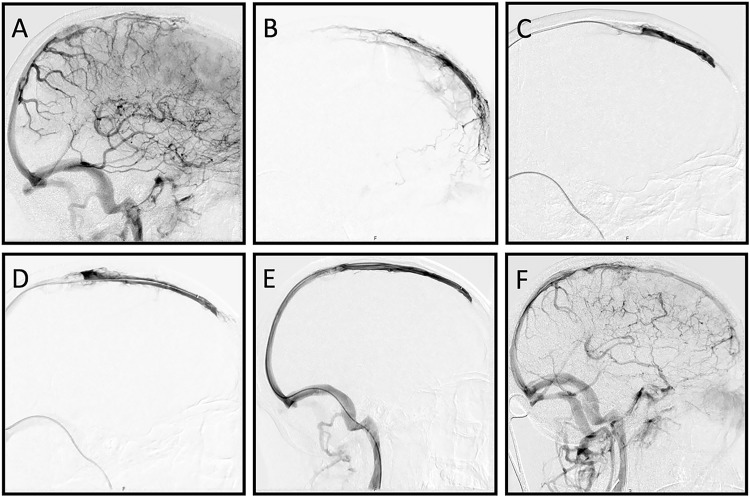
Digital subtraction angiography images, lateral views. After contrast injections in the right internal carotid artery (ICA) the venous phase confirms the occlusion of the mid to anterior part of the superior sagittal sinus (SSS) (a). A mild to moderate pseudo-phlebetic pattern of tortuous cortical veins as a response to venous congestion is also noted. Selective angiography of the SSS shows patency of the most anterior part of the SSS (b). Thrombectomy using a Trevo 6 mm × 25 mm stent retriever results in increasing recanalization after the first (c) and the second (d) stent pass. Complete recanalization is achieved after three passes (e). Contrast injection in the right ICA shows improved venous drainage through the SSS and diminished venous congestion (f).

