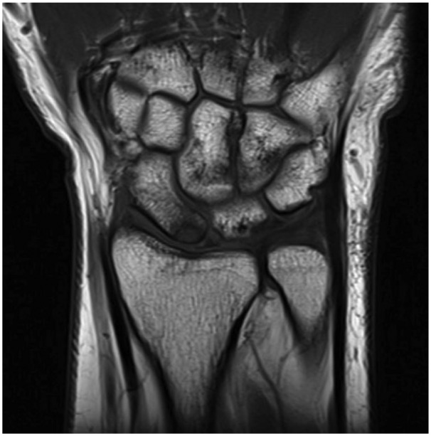
Figure 2.

Preoperative magnetic resonance imaging (T1-weighted) coronal view of the wrist illustrating the 4 × 5-mm cartilage defect along the proximal pole of the scaphoid with cartilage loss also present along the corresponding articular surface of the radius.
