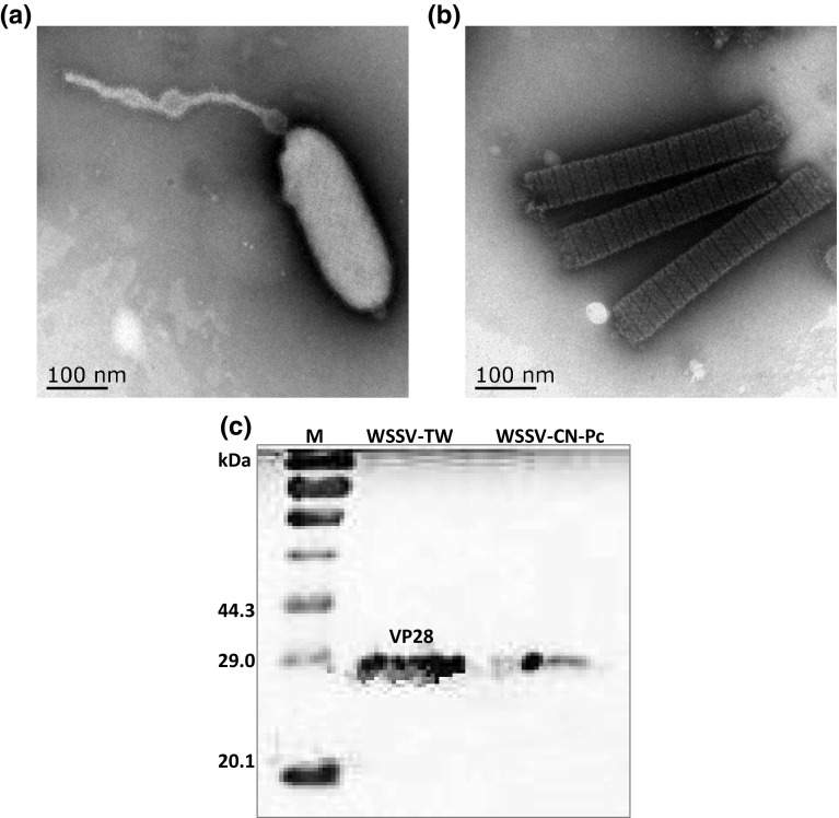
Fig. 1.
Electron micrograph and Western blot of WSSV-CN-Pc purified from crayfish samples. a Enveloped virion, scale bar 100 nm; b nucleocapsid (naked) virus (the viral envelope was peeled off due to ultracentrifugation), scale bar 100 nm; c Western blot analysis of WSSV structural protein VP28. WSSV-TW was used as positive control. M protein marker

