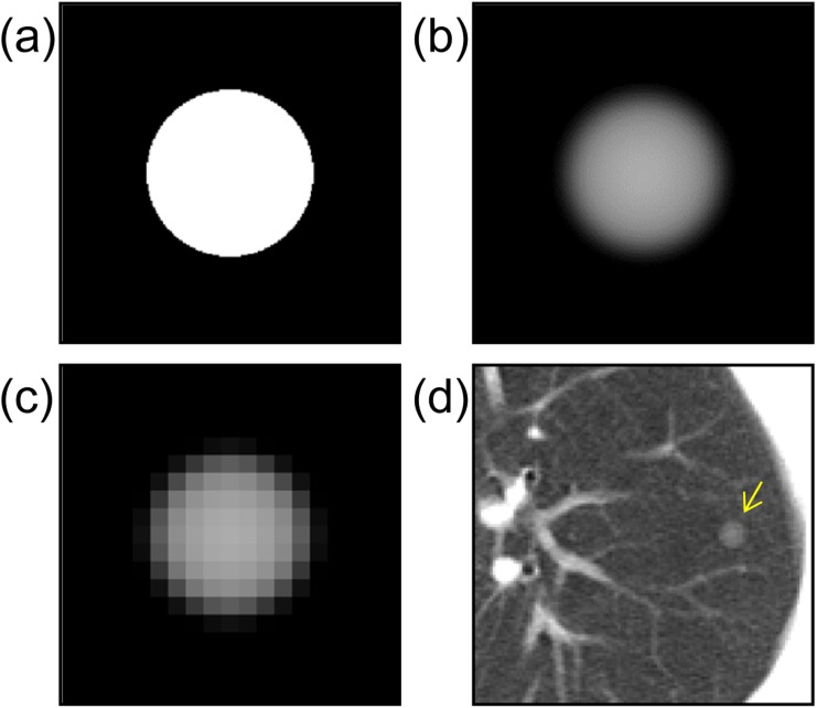
Figure 1.
A schematic explanation of virtual nodule generation: (a) the object function of a typical solitary pulmonary nodule with a diameter of 6 mm; (b) a computer-simulated nodule obtained from the object function by Equation (1); (c) a virtual nodule generated by resampling the previous image (b) in three dimensions at clinical CT image resolution; (d) a virtual nodule added to the clinical CT image (arrow).

