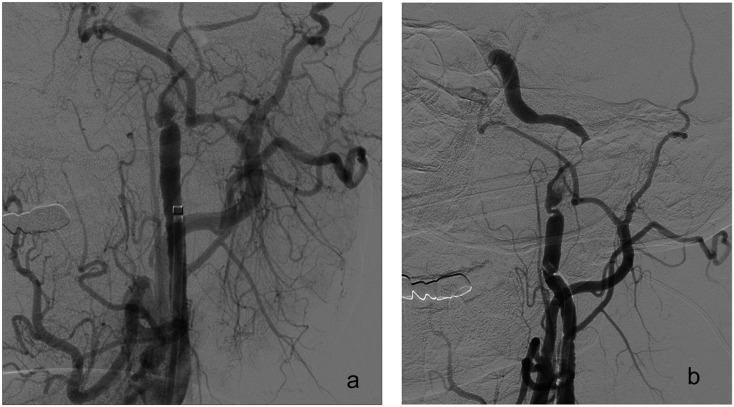
Figure 7.
(a) A digital subtraction angiography image of a 53-year-old male patient with left internal carotid artery (ICA) pseudo-occlusion is showing retarded flow at the left ICA proximal segment. (b) Left proximal ICA dissection can be observed at the left cervical ICA, while an Distal ICA bifurcation occlusion can also be spotted at the distal tip. SAT was performed to middle cerebral artery (MCA) occlusion after distal access through the dissected segment. But, the dissection caused repetitive occlusion at the ICA lumen without MCA reocclusion. Control runs from the right-side ICA showed arterial supply to the left MCA by the anterior communicating artery without delay. As a result, stenting was not performed to ICA occlusion.

