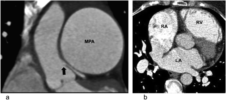
Figure 1.
Extrinsic compression of the left main stem coronary artery in a 37-year-old female with pulmonary hypertension with typical angina: (a) electrocardiogram-gated coronal CT is demonstrating extrinsic compression of the left main stem coronary artery (black block arrow) by the aneurysmal main pulmonary artery (MPA). (b) The axial CT image of the same patient is showing a sinus venosus-type atrial septal defect (white block arrow) and dilated right ventricle (RV). LA, left atrium; RA, right atrium.

