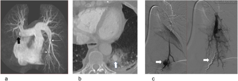
Figure 4.
Alveolar haemorrhage in a 45-year-old female with pulmonary hypertension post-right heart catheterization: (a) coronal maximum intensity projection of an MR pulmonary angiogram is demonstrating a proximal occlusion of the right middle and lower lobes (black block arrow) and proximal webs in the left lower lobe (thin white arrows) in keeping with chronic thromboembolic disease. (b) The patient developed haemoptysis following right heart catheterization. The axial CT image is showing a pseudoaneurysm of the posterior basal left lower lobe artery (white block arrow) and surrounding alveolar haemorrhage that developed as a consequence of forced passage of the catheter across the intraluminal webs. (c) Selected images of catheter pulmonary angiography before (left) and after (right) embolization of the left lower lobe pseudoaneurysm (block white arrows).

