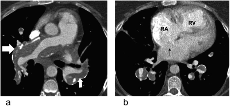
Figure 5.
In situ thrombus in a 57-year-old female with long-standing pulmonary hypertension secondary to congenital heart disease: (a) the axial non-electrocardiogram-gated CT pulmonary angiogram with calcified thrombus lining the proximal pulmonary arteries (white block arrows). (b) There is dilatation of the right atrium (RA) and right ventricle (RV) with right ventricular hypertrophy and an ostium secondum atrial septal defect (black thin arrow). The segmental vessels are large and contain partially occlusive calcified thrombus.

