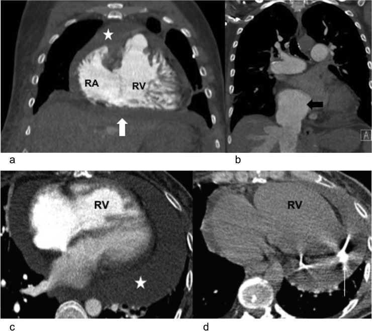
Figure 6.
Right heart failure and pericardial tamponade in two different patients with pulmonary hypertension: (a) coronal CT pulmonary angiogram is demonstrating dilatation of the right ventricle (RV) and hypertrophy with pericardial effusion (star) and ascites (block white arrow). There is also mild subcutaneous oedema. (b) The same patient also had a dilated inferior vena cava (block black arrow) and distended hepatic veins. (c, d) Axial CT pulmonary angiogram in a different case is demonstrating right ventricular dilatation and hypertrophy with a moderately large circumferential pericardial effusion (star). Clinically, the patient had signs of tamponade. A CT-guided pericardial drain (thin white arrow) was inserted with good improvement in symptoms. RA, right atrium.

