
Figure 3.
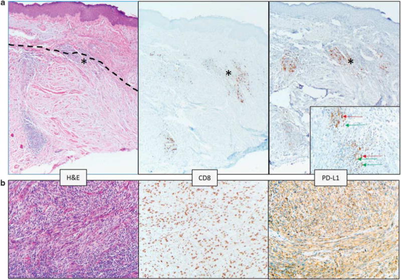
Pure desmoplastic melanomas demonstrate a different PD-L1 expression pattern than other CSD melanomas. (a) The dotted line in the left panel delineates the upper boundary of a desmoplastic melanoma centered in the dermis. Solar elastosis is a prominent feature in the superficial dermis. Lymphoid aggregates are present, and one at the boundary of the melanoma and normal dermis is marked with an asterisk (*) and is shown on the inset of the right panel. In the pure desmoplastic melanomas, PD-L1 expression was observed on lymphocytes (green arrow on inset) and macrophages (red arrows on inset) in lymphoid aggregates. This case did not show PD-L1 expression on tumor cells. Original magnification, ×100, inset ×400. (b) CSD melanoma with spindled morphology showing 60% tumor cell PD-L1 expression associated with a ‘severe’ grade CD8+ TIL infiltrate. ‘Severe’ grade is defined as a diffuse infiltrate of TIL throughout the melanoma. ×200 original magnification.