
Figure 4.
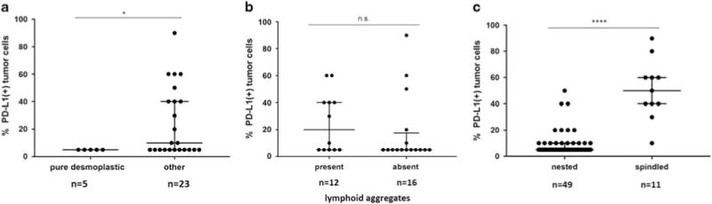
Association of morphologic features with levels of PD-L1 expression. (a) Among PD-L1(+) CSD melanomas, there were significantly higher median PD-L1 expression levels in cases lacking a pure desmoplastic phenotype (*P = 0.047, Mann–Whitney U-test). (b) The presence or absence of lymphoid aggregates did not correlate with the levels of PD-L1 expression observed in PD-L1(+) CSD melanomas (P = 0.22). (c) Among all melanoma subtypes, PD-L1(+) tumors with a spindled morphology demonstrated a higher percentage of tumor cells with PD-L1 display compared to those with a nested morphology (****P<0.0001). n.s., not significant.