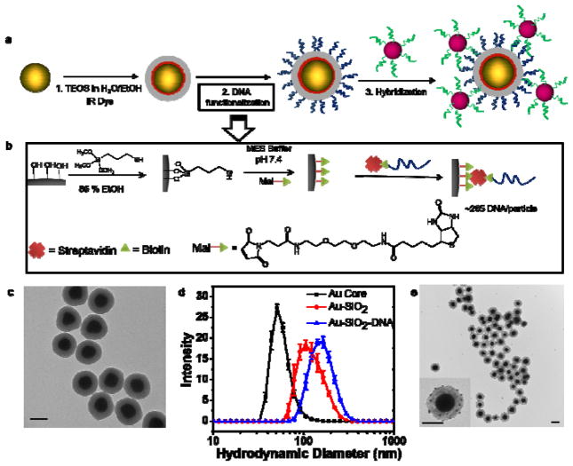
Figure 2.
(a) Synthesis and DNA functionalization of SERS NPs. First, 70 nm spherical AuNP cores were silica coated in the presence of IR dye (IR 780 perchlorate or IR 792 perchlorate) to produce SERS NPs. The SERS NPs were functionalized with DNA molecules by sequential modification of the silica shell surface. To verify the presence of the DNA sequence, a 10 nm AuNP functionalized with the complementary DNA strand was hybridized to produce SERS NP core - 10 nm AuNP satellite hybrid nanostructures. (b) Illustration of DNA functionalization: first, the surface hydroxyl groups were converted to sulfhydryl groups using (3-mercaptopropyl)trimethoxysilane (MPTMS) in a water-ethanol mixture. Next, sulfhydryl groups were reacted with a maleimide-PEG2-biotin linker to attach biotin molecules to the surface. Subsequently, biotin labeled DNA molecules were attached to the biotinylated surface of the SERS NPs using neutravidin, a biotin-binding protein as a crosslinker. (c) Representative TEM images of the DNA functionalized SERS NPs. (d) The DLS hydrodynamic diameter measurements show an increase in size from ~80 nm to ~137 nm after silica coating and ~170 nm after DNA functionalization. Error bars represent standard deviations of three measurements. (e) Representative TEM images of the SERS NP core – 10nm AuNP satellite hybrid nanostructures. The inset shows 10 nm AuNP satellites being attached to the SERS NP surface. All the scale bars are 100 nm.

