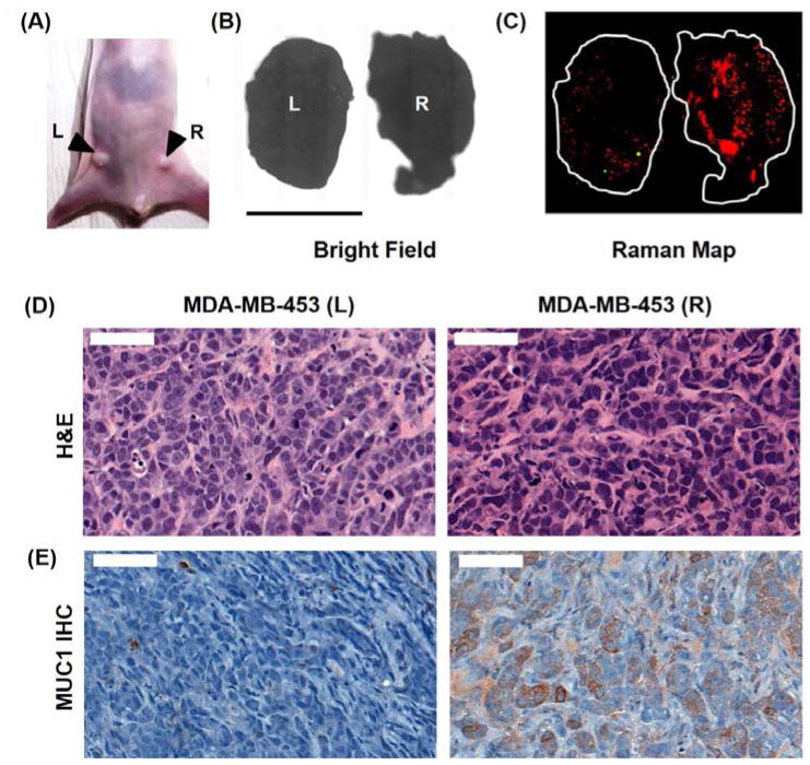
Figure 6.
(a) Photograph of a nude athymic mouse with MDA-MB-468 tumor (R) and MDA-MB-453 (L) xenograft. (b) Bright field image of the excised tumors. (c) Ex vivo Raman image of the tumors (100% laser power, 1.5 s integration time, 5× objective). The predominantly red signal corresponds to the prevalence of MUC1-NPs throughout the tumor volume. On the other hand, the MDA-MB-453 (L) tumor showed minimal uptake of both, MUC1-NPs and NT-NPs. (d) Hematoxylin and eosin (H&E) staining of the cancer tissue. (e) Immunohistochemical (IHC) staining of MUC1 in the fixed tumor tissue demonstrating overexpression of MUC1 in the MDA-MB-468 tumor in contrast to the MDA-MB-453 tumor.

