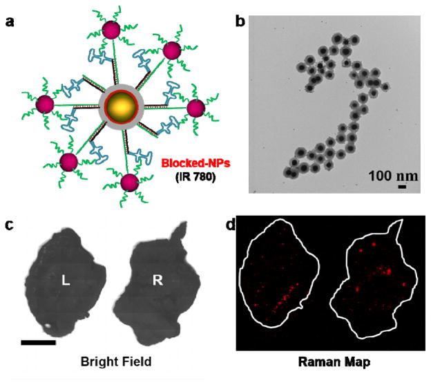
Figure 7.
(a) Schematic representation of the blocked MUC1-NPs. The aptamer moiety is sterically shielded by 10 nm AuNPs. (b) TEM images of the blocked MUC1-NPs after 16 hours of serum incubation at 37 °C showing excellent structural integrity of the hybrid structures. (c) Bright field image of the excised tumors. (d) Ex vivo Raman image of the tumor (100% laser power, 1.5 s integration time, 5× objective). Both tumors exhibited very low uptake of the blocked NPs, suggesting that NP homing is due to the active targeting of the aptamers grafted on the MUC1-NPs.

