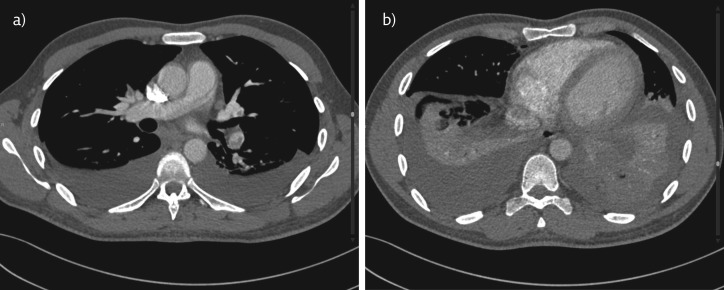
Figure 4.
a) CTPA showing filling defects in both the upper and left lower lobar pulmonary arteries suggestive of bilateral pulmonary embolism. Bilateral, mild pleural effusion is also seen. b) CTPA showing no enlargement of the right ventricle with a normal position of the interventricular septum suggesting no evidence of right ventricle dysfunction.

