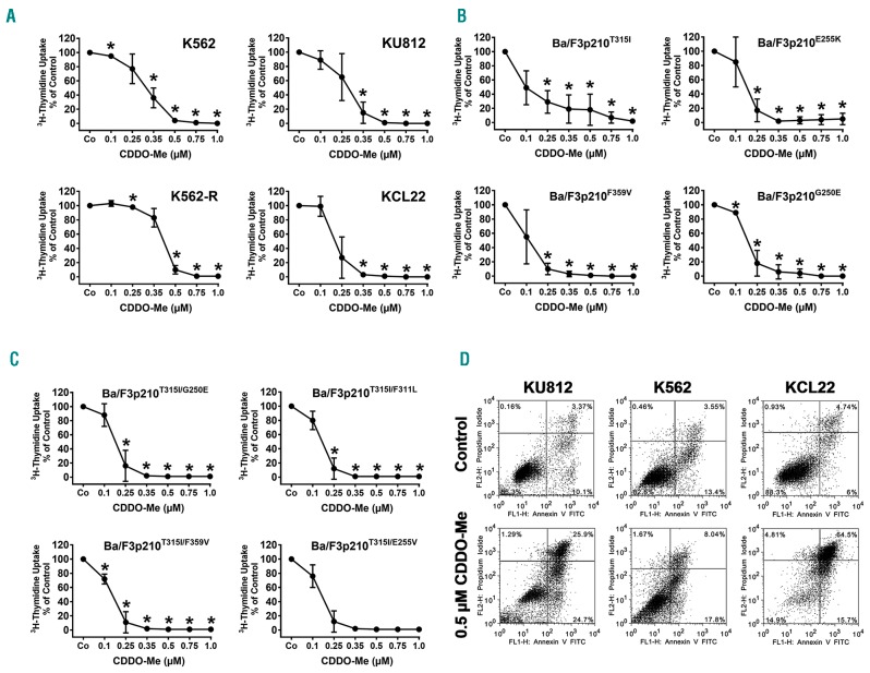
Figure 1.
CDDO-Me inhibits growth and viability of Philadelphia-positive (Ph+) cell lines. (A–C) Human chronic myeloid leukemia (CML) cell lines (A) and BCR-ABL1-expressing Ba/F3 cells (B and C) were exposed to control medium (Co) or various concentrations of CDDO-Me for 48 hours (h). In case of K562-R, imatinib was removed prior to CDDO-Me exposure. Then, proliferation was measured by assessing 3H-thymidine uptake. Results are expressed in % of control and represent the mean±Standard Deviation (S.D.) of 3 independent experiments. (D) CML cell lines (K562, KU812, KCL22) were incubated in control medium or in CDDO-Me (0.5 μM) for 48 h. Thereafter, the percentage of apoptotic cells was determined by combined Annexin V/PI staining. The figure shows dot blots from one representative experiment. Almost identical results were obtained in two other experiments.

