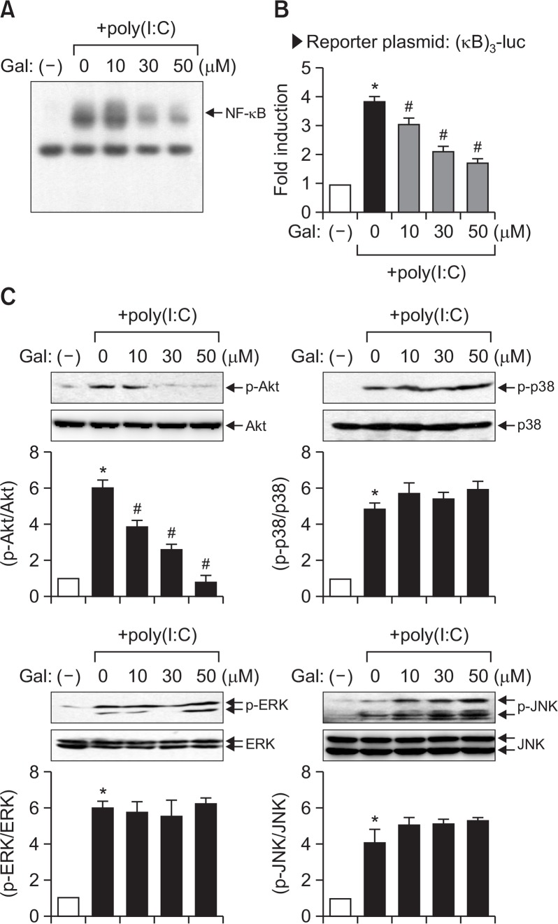
Fig. 4.
Effect of galangin on NF-κB activity and the phosphorylation of Akt and MAP kinases in poly(I:C)-stimulated BV2 cells. (A) Nuclear extracts were prepared from BV2 cells after treatment with poly(I:C) (10 μg/ml) for 3 h, in the absence or presence of galangin, and EMSA was performed to determine the DNA binding activity of NF-κB. The arrow indicates the DNA-protein complex of NF-κB. (B) BV2 cells transfected with the reporter plasmid ([κB]3-luc) were pretreated with galangin for 1 h prior to poly(I:C) treatment. After 6 h, cells were harvested and a luciferase assay was performed. (C) Western blot analysis for MAPKs and PI3K/Akt activities. Cell extracts were prepared from BV2 cells treated with poly(I:C) for 30 min, in the absence or presence of galangin, and a western blot analysis was performed using antibodies against phospho- or total forms of three types of MAPKs or Akt. The blots are representative of three independent experiments. Quantification of data from western blot analysis is shown in the bottom panel. The levels of the phosphorylated forms of MAPKs or Akt were normalized to the levels of each total form and expressed as relative fold changes versus the untreated control group. The results were obtained from three independent experiments and expressed as the mean ± SEM. *p<0.05, vs. control samples. #p<0.05, vs. poly(I:C)-treated samples.

