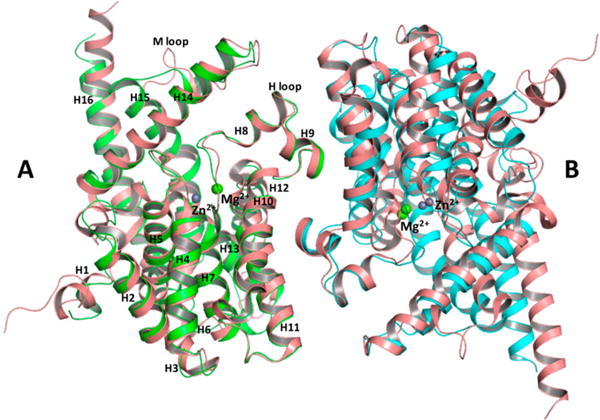
Figure 10.

Superposition of PDE and PDE9 (PDB entry 2HD1) dimers using monomer A as a reference molecule. Monomers A and B of PDE are colored green and cyan, respectively. Both monomers of PDE9 are colored salmon. Zn2+ (purple) and Mg2+ (green) ions are modeled as spheres. Helices of monomer A are labeled according to the PDE9 model.
