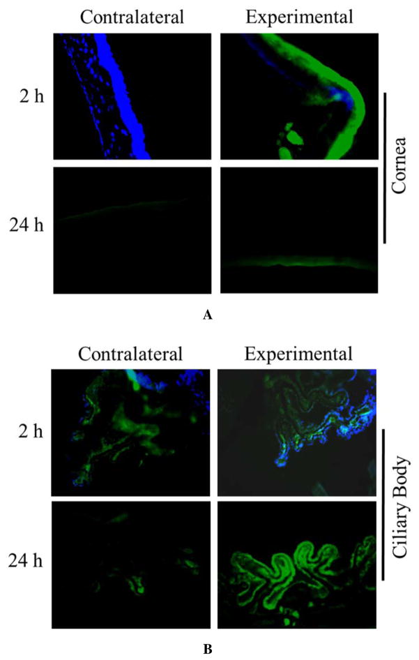
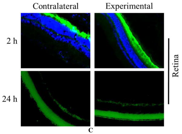
Figure 7. Ocular disposition of DNF.
Brown Norway rats (n=3) received a DNF-FITC mat topically in the right eye (experimental eye) while the left eye received no treatment (contralateral eye). Animals were euthanized at 2 or 24 h and the ocular tissues harvested and immediately processed for cryosectioning. Fluorescent imaging of sections from various ocular organs showed most dendrimers were flushed from the cornea in 24 h. Over the same time period, FITC-G3.0-mPEG accumulated in the ciliary body of the experimental eye.


