FIG. 2.
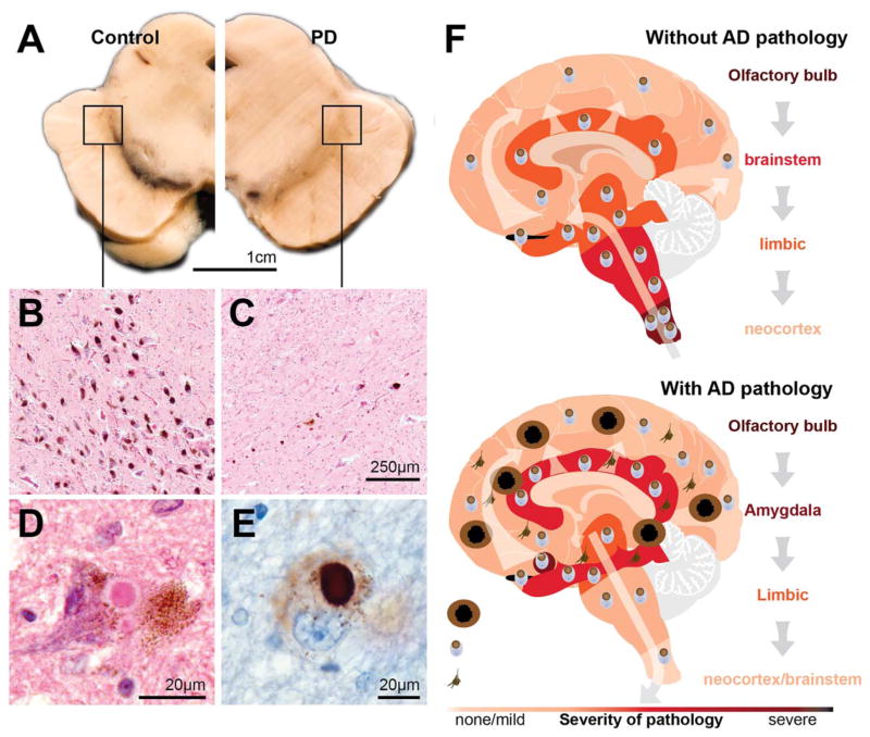
The main pathologies in patients with clinical Parkinson’s disease and the pathological progression. (A) Transverse hemisection of the midbrain of a control at left and a patient with clinical Parkinson’s disease (PD) at right showing the marked reduction in the black pigment within the substantia nigra region. (B–C) Haematoxylin and eosin stained section of the ventrolateral region identified by the box in A showing at higher magnification the pigmented neurons of the substantia nigra in a control without PD (B) and a person with clinical PD (C). (D–E) Intracytoplasmic Lewy bodies in remaining pigmented neuron of the substantia nigra of a patient with PD showing the eosinophilic core and paler halo in haematoxylin and eosin stain (D) and the dark aggregation of α-synuclein using immunoperoxidase with cresyl violet counterstaining (E). (F) Cartoon representation (based on data from Toledo et al73) of the two major patterns of Lewy-body pathology in patients with (below) and without (above) Alzheimer’s disease (AD) pathology. In those with clinical PD and little AD pathology, Lewy bodies begin in the olfactory bulb and medulla oblongata then infiltrate higher brain stem regions, then limbic brain regions, and finally the neocortex. In those with AD pathology, this pattern is different. Lewy bodies concentrate in limbic regions of the brain prior to infiltrating to other regions.

