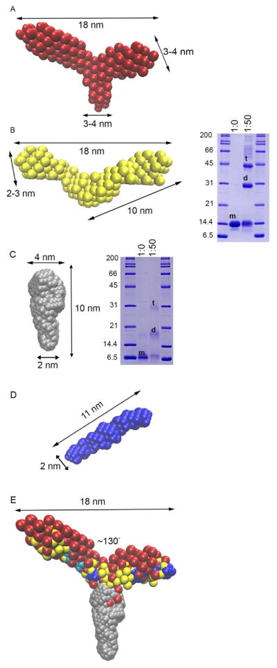
Figure 3. Ab initio models from SEC-SAXS reveal tripartite structure for myocilin CC.

(A) CC60-185 is Y-shaped with arms spanning 18 nm and 130° apart from a stem ~3–4 nm in width. (B) Left: CC69-185 is a P2-symmetric V-shaped molecule with similar dimensions to arms of CC60-185. Right: Chemical crosslinking of CC69-185 reveals both dimer and tetramer species. m=monomer, d=dimer, t=tetramer. (C) Left: CC33-111 is an oblong molecule with varying widths of 2–4 nm. Right: Chemical crosslinking of CC33-111 indicates dimer and tetramer species with labels as in (B). (D) CC112-185 is a P2-symmetric, ~11 nm long rod. (E) Overlay of SAXS molecular envelopes from A–D. Molecular envelopes are overlaid on the same scale. Two orientations for CC112-185 are shown in dark and light blue. See also Figures S2, S4, and Table 2.
