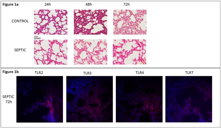
Fig 1.
a) Lung tissue sections are stained with hematoxylin and eosin. Images (x40) from lung sections are shown at 24h, 48h and 72h after sham operation or CLP procedure. Control mice (sham operated), (upper panel in 1a) and septic mice (down panel in 1a) demonstrate no lung injury and increased lung injury observed during time period, respectively. Different degrees of acute lung injury assessed by histological examination in septic mice. Observed signs of edema and alveolar damage detected from 24 h after CLP, including alveolar flooding and alveolar collapse. b) Immunofluorescence staining expressing TLR 2, TLR 3, TLR 4 and TLR 7 in capillaries at 72h after CLP challenge. Among septic mice, the highest expression of TLRs was noted at this time point (down panel). TLR-positive cells in the lung of septic mice were stained red, while cell nuclei were stained blue. Original magnification x63.

