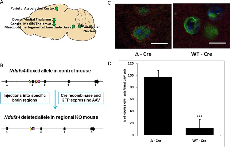
Fig 1. Viral injections and characterization.
A. Schematic diagram depicting the five regions which were injected with either rAAV/WT-Cre GFP (knockout virus expressing functional Cre recombinase) or rAAV/Δ-Cre GFP (sham virus expressing nonfunctional Cre recombinase). B. Schematic showing the mechanism of gene deletion. Exon 2 of genomic Ndufs4 is excised by the virus expressing active Cre recombinase, resulting in the loss of NDUFS4 protein as described by Kruse et al. [6]. C. Loss of Ndufs4 expression in active Cre virus infected cells in the PAC. Infected cells appear green under the confocal microscope due to the viral GFP co-expression (Magnification X1000). In the Δ-Cre sham virus infected cells of the PAC, mitochondrial NDUFS4 fluorescence (red) is seen, which is absent in the WT-Cre infected cells. D. Representative quantitation of NDUFS4 expression in the virus infected cells, shown as a percentage of total infected cells. 20 image fields were quantified per injected locus. Scale bar: 10μm. *** indicates p-values <0.001.

