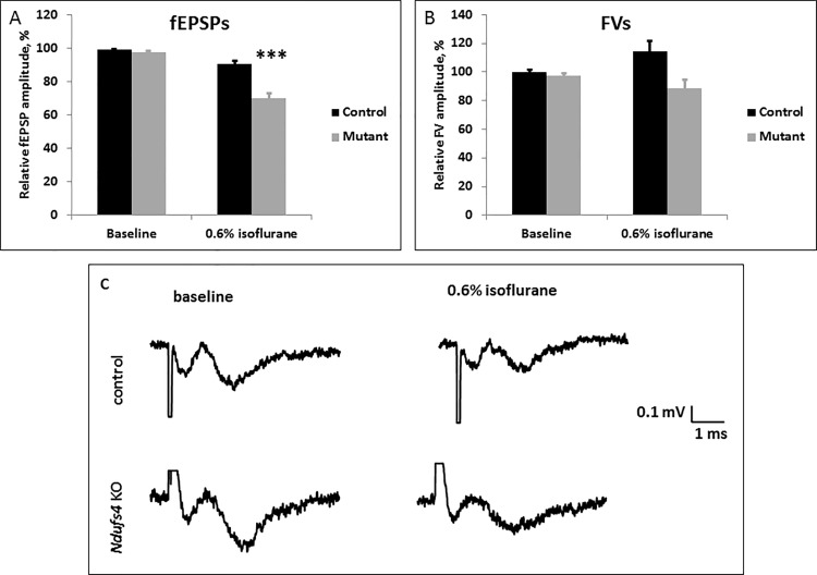
Fig 8. Electrophysiological field recordings of the excitatory synaptic transmission.
A. Amplitudes of fEPSPs decreased with 0.6% (248 μM) ISO exposure in control and Ndufs4(KO) brain slices. The decrease of fEPSPs was significantly greater for the Ndufs4(KO) than the control (*** indicates p-value <0.001). B. Fiber volleys of both genotypes were not significantly affected by the isoflurane exposure. C. Representative traces of field recordings from control and KO slices before and during exposure to 0.6% ISO (equivalent to 248 μM). 0.6% ISO exposure led to a significantly larger decrease in the amplitudes of fEPSP in the KO when compared to the respective decrease in controls. The amplitudes of the fiber volleys were decreased (statistically tending to significance, p = 0.015) by 0.6% ISO exposure in both genotypes.

