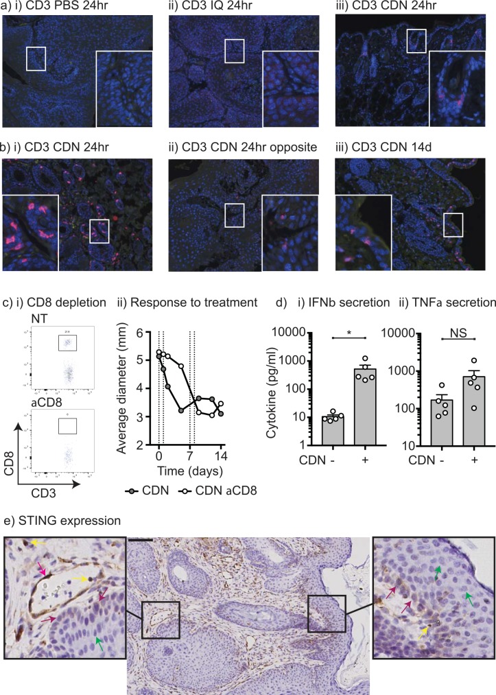
Fig 2. Infiltration of T cells following treatment with STING ligand.
Pdx-Cre+/- Kras(G12D)+/- Trp53(R172H)+/- mice exhibiting papilloma were injected with i) PBS vehicle, ii) 25μg Imiquimod, or iii) 25μg CDN and the site harvested 24 hours later for histology. Sections were stained for CD3 (pink) and DAPI nuclear counterstain (blue). Insets are an enlargement of regions of interest. b) A Pdx-Cre+/- Kras(G12D)+/- Trp53(R172H)+/- mouse exhibiting dual papilloma on opposite sides of the face was injected with 25μg CDN to one lesion and the other left untreated. 24 hours later both papilloma sites were harvested and stained for CD3 (pink) and DAPI nuclear counterstain (blue) on the i) treated and ii) untreated opposite side papilloma. iii) CD3 (pink) and DAPI nuclear counterstain (blue) in a CDN-treated papilloma 14 days following initiation of treatment. Insets are an enlargement of regions of interest. c) Pdx-Cre+/- Kras(G12D)+/- Trp53(R172H)+/- mice exhibiting papilloma were left untreated or depleted of CD8 T cells with 50μg anti-CD8 ip. 1 day prior to treatment with 25μg CDN injected on d0, 1, 7 and 8. Detection of CD3+CD8+ T cells in the peripheral blood on d0 in representative i) NT or ii) CD8-depleted mice. iii) Average size of papilloma through treatment. d) Explants of papilloma were left untreated or treated with 25μg/ml CDN overnight, and supernatants were assessed for section of i) IFNβ and ii) TNFα by multiplex bead assay. e) Immunohistology for STING expression in Pdx-Cre+/- Kras(G12D)+/- Trp53(R172H)+/- murine papilloma. Insets are an enlargement of regions of interest. Arrows depict STING+ endothelial cells (red arrows), STING+ immune cells (yellow arrows), STING+ basal cells (purple arrows), and STING- differentiated non-basal epithelial cells (green arrows).

