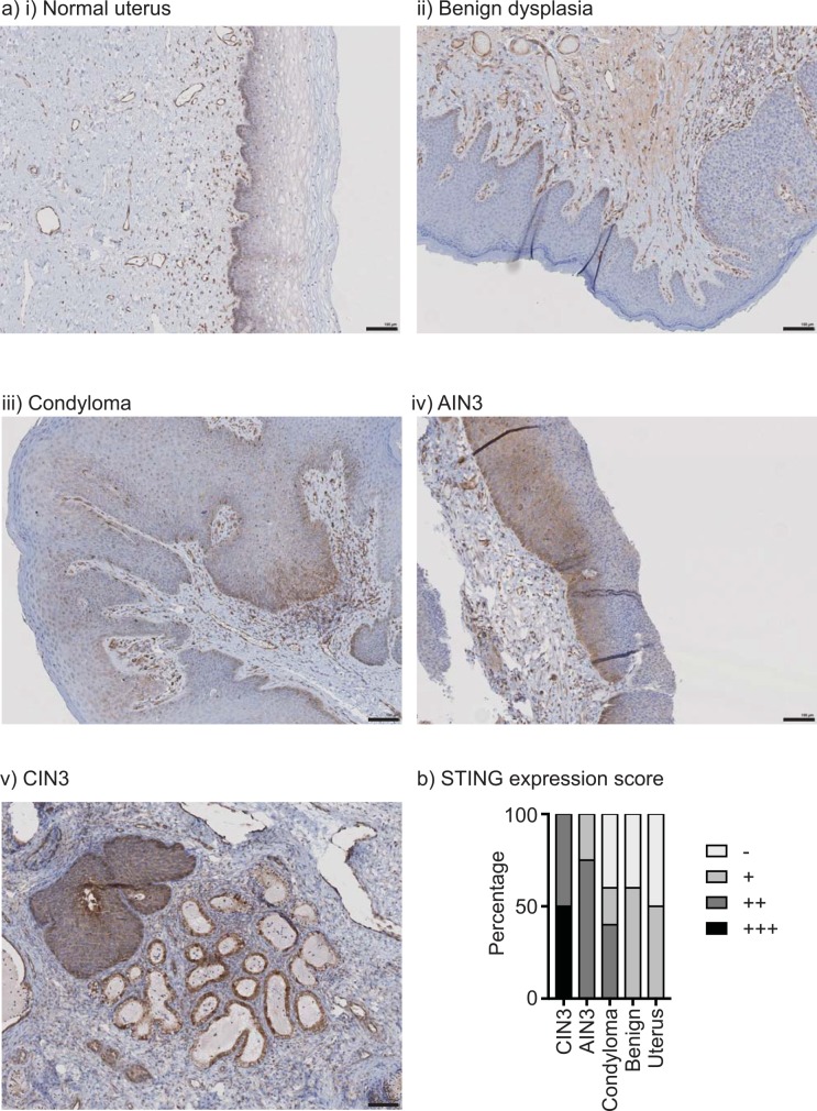
Fig 4. STING expression in HPV-associated disease.
a) Examples of STING staining in i) normal uterus, ii) benign dysplasia, iii) condyloma, iv) AIN3, v) CIN3. b) The degree of STING staining was scored on a scale from negative (-) to highly positive (+++). The graph shows a summary of the proportion of each histology with each staining pattern.

