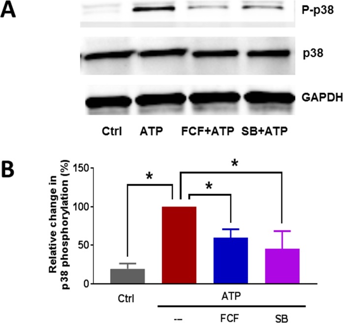
Fig 4. eATP induced phosphorylation of p38 MAPK in HSV endothelial cells.
HSVEC were untreated (Ctrl), or treated with ATP (2 mM) for 30 minutes. Some cells were pre-treated with FCF (100 μM), or SB203580 (SB, 20 μM) for 1 hour. Phospho-p38 MAPK and total p38 MAPK proteins were quantitated with immunoblotting (adjusted to the loading control GAPDH). (A) Representative western blots of phospho-p38 MAPK and p38 MAPK. (B) Cumulative data showing the relative percent phosphorylation of p38 MAPK. Phosphorylation with eATP alone was set as 100%, n = 4 passages,* p < 0.05, (One way ANOVA).

