
Figure 6.
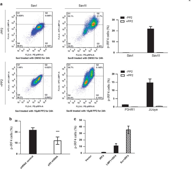
The level of IRF4 phosphorylation correlates with LMP1 and is dampened by the Src-specific inhibitor PP2 in EBV+ cells. (a) Cells were collected, washed and fixed after treatment with 15 μM PP2 or DMSO (control) for 24 h. Cells were then permeabilized, and incubated with mouse anti-LMP1 antibody (Dako) and rabbit anti-p-IRF4 antibody (21st Century Biochemicals). After wash, cells were incubated with anti-mouse IgG APC (eBioscience) and anti-rabbit PE (eBioscience) before subjected to flow cytometry analysis. Q4 represents the portion of LMP1+/p-IRF4+ cells. (b) SavIII cells were transfected with p85 shRNA (#1) (or control). shRNA expression was induced by 1 μg/ml doxycycline for 3 days. p-IRF4 was then evaluated by flow. P = 0.0007 (unpaired t-test). (c) BJAB cells were transfected with IRF4, IRF4 plus LMP1 (or c-Src as a positive control). Cells were subjected to flow analysis after 48 h. Statistical analysis was performed for three independent flow analyses. Results are the averages ± s.d. Representative results from at least three independent experiments are shown.