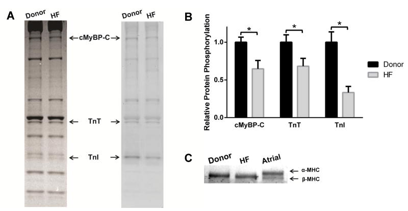
Figure 1. Phosphorylation of sarcomeric proteins and MHC expression in donor and HF myocardium.
(A) Representative Coomassie stained (left) SDS gel and Pro-Q Diamond-stained (right) showing the expression and phosphorylation status of myofilament proteins in donor and HF samples. Cardiac samples isolated from 4 donor hearts and 4 failing hearts were used to analyze contractile protein expression and phosphorylation levels. (B) Quantification of phosphorylation of MyBP-C, TnT, and TnI in donor and HF samples. (C) Representative 5% Tris-HCl gel showing MHC isoform expression in donor and HF myocardium. Values are expressed as mean ± S.E.M. * P < 0.05.

