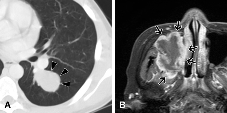
Figure 3. Imaging of inflammatory myofibroblastic tumors.
A. Chest computed tomography reveals a tumor with smooth and well-defined margins in the lung parenchyma of a 12-year-old boy (Case 2). This tumor is positive for ALK rearrangement. B. Head magnetic resonance imaging reveals a lobulated irregular tumor arising in the sinusoidal space of a 70-year-old woman (Case 24). This tumor is negative for ALK rearrangement. Resection was incomplete during the first surgery, and the patient experienced local recurrence after 1 month.

