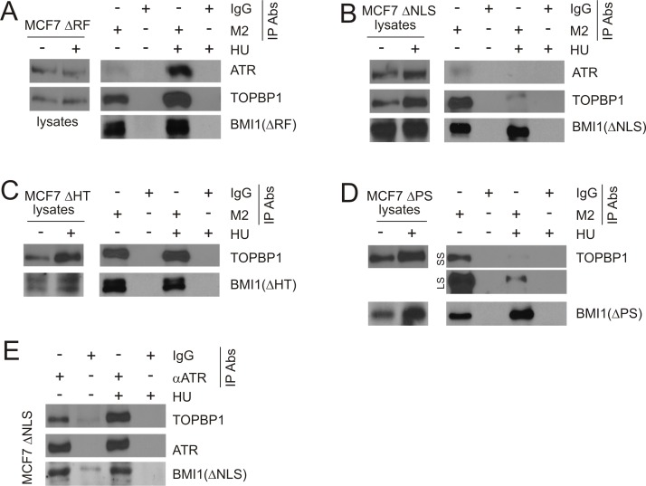
Figure 8. Characterization of BMI1 association with TOPBP1 and ATR.
Cell lysate from the indicated MCF7 BMI1ΔRF, BMI1ΔNLS, BMI1ΔHT, or BMI1ΔPS cells (A–E) were pre-treated with benzonase at 1 U/μl on ice for 1 hour, followed by IP with a monoclonal anti-FLAG (M2) (A–D) or anti-ATR antibodies (E). Western blot analyses were performed for ATR, TOPBP1, and the indicated BMI1 mutants (using a polyclonal anti-FLAG antibody). 1/10 of cell lysates used for IP were also analyzed. Experiments were repeated at least three times; typical results from a single repeat are shown. (D) LS: long exposure; SS: short exposure.

