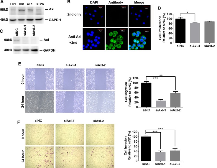
Figure 1. The expression and biological role of mouse Axl in mouse tumor cells.
(A) Axl expression in 4 mouse tumor cell lines was determined by Western blotting. Data are representative of two independent experiments. (B) Axl expression in ID8 cells was evaluated by immunofluoresecent staining. Up and bottom pictures display the control (only 2nd staining) and Axl staining respectively. 2nd: secondary antibody; Scale bar, 30 μm. (C) Efficient knockdown of Axl expression in ID8 cells transfected with control (siNC) or siRNA specific for Axl (siAxl-1 and siAxl-2). (D) The proliferation of ID8 cells with or without Axl knockdown. Proliferation was plotted as a percentage of growth relative to control siNC-treated cells. (E) ID8 cells with or without Axl knockdown were subjected to wound exposure. Wound length was imaged and measured after the wound was first made (0 hour) and 24 hours post wound exposure with statistics and representative images (10× magnification) shown. Migration was plotted as a percentage relative to zero time point of each treated cells. (F) ID8 cells with or without Axl knockdown were subjected to invasion assay with statistics and representative images (10× magnification) shown. Invasion was plotted as a percentage relative to control siNC-treated cells. The experiments were performed twice with 3 technical replicates, and data are expressed as mean ± SEM (for D–F), *p < 0.05, **p < 0.01, ***p < 0.001, one-way ANOVA followed by Tukey's multiple comparisons test.

