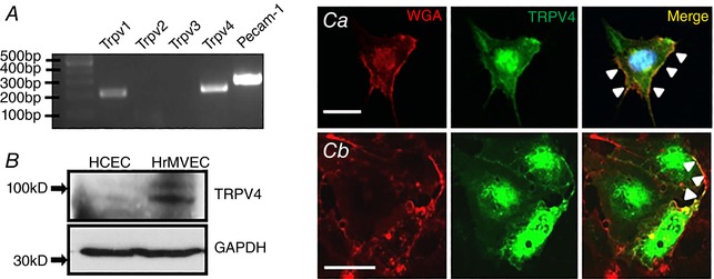
Figure 1. TRPV4 is expressed in retinal microvascular cells.

A, RT‐PCR. Trpv1, Trpv4 and Pecam‐1 but not Trpv2 & 3 transcripts are expressed in HrMVECs. B, Western blot. TRPV4 protein expression is prominent in HrMVECs but weak in HCECs. C, HrMVEC labelled by anti‐TRPV4 antibody and a plasma membrane marker (WGA; wheat germ agglutinin) (Ca). The TRPV4 signal within the plasmalemma is marked by arrowheads. Transfection with the Trpv4:eGfp construct (Cb). While TRPV4 expression is predominantly cytosolic, the TRPV4‐ir signal colocalizes with WGA at the plasma membrane (arrowheads). Scale bar = 50 μm. [Color figure can be viewed at wileyonlinelibrary.com]
