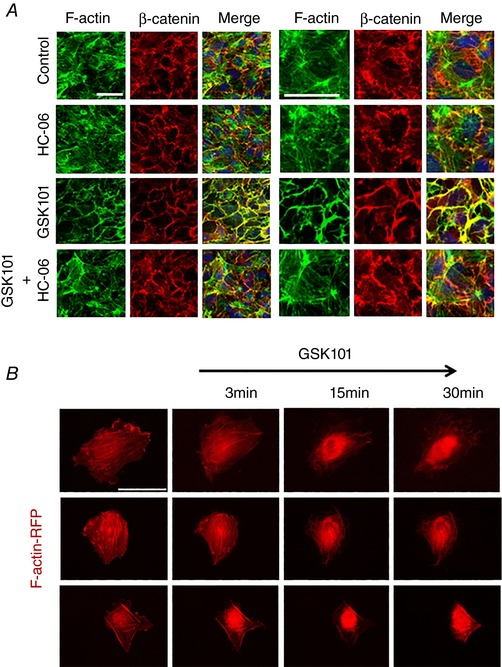
Figure 7. TRPV4‐dependent remodelling of F‐actin and AJs in confluent HrMVECs.

A, double labelling for β‐catenin and F‐actin. Combined images include DAPI staining (blue). GSK101‐induced disruption of central and cortical F‐actin was rescued by the TRPV4 antagonist (HC‐06). Scale bar = 50 μm. B, red fluorescent protein (RFP)‐actin‐transfected HrMVEC cells exposed to GSK101 (5 nm) show time‐dependent redistribution of F‐actin towards the cell nucleus. Scale bar = 50 μm. [Color figure can be viewed at wileyonlinelibrary.com]
