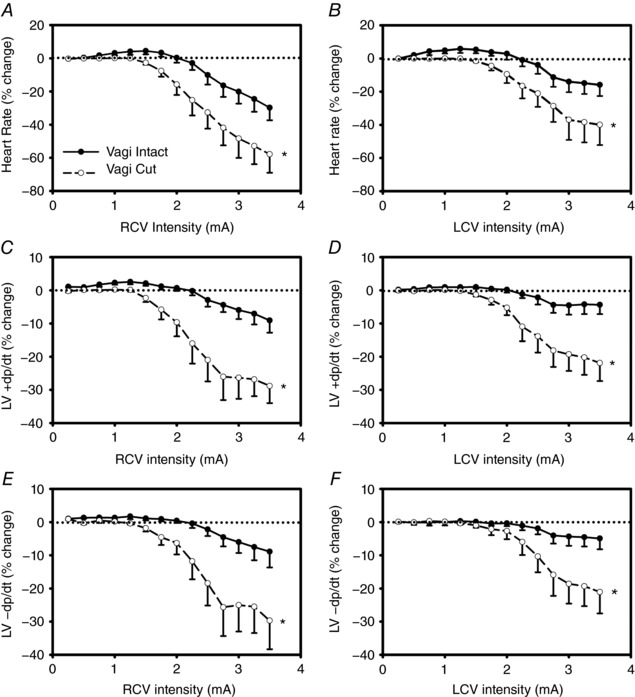
Figure 9. Evoked changes in chronotropic (A and B), left ventricular inotropic (C and D) and lusitropic (E and F) function in response right (left panels) and left (right panels) cervical VNS prior to and following cervical vagus transections rostral to stimulating electrode.

Animals were anaesthetized throughout. VNS delivered at 500 μs pulse width and a 17.5% duty cycle with a 14 s on‐phase. Responses reflect percentage change from baseline during VNS as a function of stimulus frequency. * P < 0.001 vs. intact.
