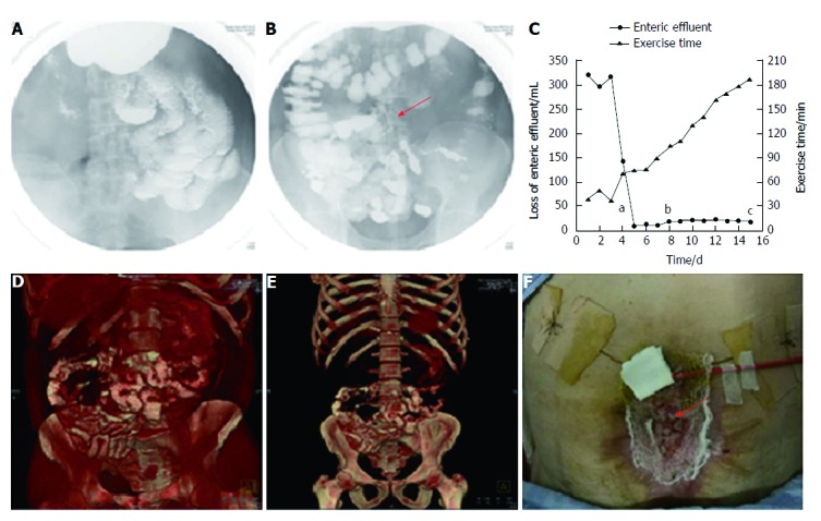
Figure 3.

Follow-up after stent implantation. A: Image of small intestine after barium meal, indicating no obstruction; B: Image of colon after barium meal, indicating that contrast passed through the stent smoothly (red arrow: fistula stent); C: Loss of enteric effluent and the time of exercise tolerance before and after stent implantation. aTime point of stent insertion; bTime point of starting EN; cTime point of skin transplantation; D and E: 3D-reconstructed images after barium meal, indicating no abnormity after starting EN; G: Skin transplantation for abdominal wall reconstruction (red arrow: skin graft). EN: Enteral nutrition.
