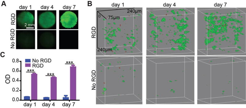
Figure 3.

3D culture of HUVECs in RGD-functionalized porous PEG hydrogel. A) Whole hydrogel imaging. HUVECs were stained with Calcein AM. B) Confocal microscopy images of HUVECs in the porous hydrogels. HUVECs were stained with the Live/Dead viability kit. The green color represents live cells and the red color represents dead cells. C) MTS test of HUVEC proliferations. ***, p < 0.001.
