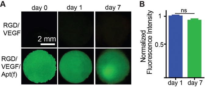
Figure 6.

Staining of aptamers in the hydrogel. A) Imaging of the whole hydrogels treated with an FAM-labeled complementary sequence after the hydrogels were incubated in the cell culture media with cells for 0, 1, and 7 d. RGD/VEGF: RGD-functionalized VEGF-loaded hydrogel. RGD/VEGF/Apt(f): RGD/aptamer-functionalized VEGF-loaded hydrogel. B) Fluorescence intensity of the RGD/VEGF/Apt(f) hydrogels at day 1 and 7. The values were normalized to that of day 0. ns: not significant.
