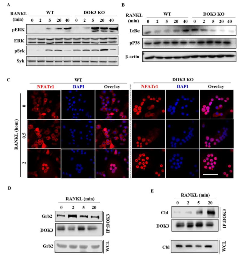
Figure 3. DOK3 negatively regulates RANKL signaling.

(A) WT and DOK3-/- BMMs were stimulated with RANKL (100 ng/ml) for the indicated times. Cell lysates were analyzed by Western blotting for phosphorylated and total Syk and ERK. (B) WT and DOK3-/- pre-osteoclasts were stimulated with RANKL (100 ng/ml) over time. Cell lysates were immunoblotted for IκBα, phosphorylated p38, and β-actin. (C) WT and DOK3-/- BMMs were stimulated with RANKL (20 ng/ml) for two days to induce NFATC1, serum starved for 4 hours, and stimulated with RANKL for indicated times to induce nuclear translocation of NFATc1. Cells were fixed, permeabilized, and NFATc1 localization detected by confocal microscopy. (D and E) BMMs were stimulated with RANKL (100 ng/ml) over time, cell lysates were immunoprecipitated with anti-DOK3 antibody and co-precipitated proteins analyzed by Western blot for Grb2 (D), Cbl (E) and DOK3. Scale bar: 200 μm (C). Experiments was repeated three times.
