
Figure 4. DOK3 limits M-CSF induced signaling and proliferation of pre-osteoclasts.
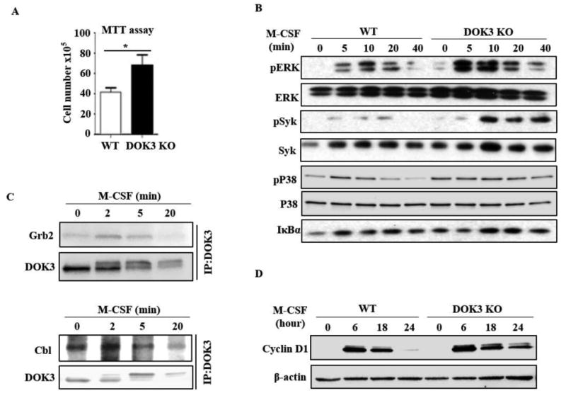
(A) WT and DOK3-/- BMMs were stimulated with M-CSF 100 ng/ml for 24 hours followed by MTT reduction assay. (B) WT and DOK3-/- BMMs were stimulated with M-CSF (100 ng/ml) for the indicated times and cell lysates were immunoblotted for phosphorylated and total ERK, Syk, and P38, and IκBα. (C) BMMs were stimulated with M-CSF (100 ng/ml), cell lysates were immunoprecipitated with anti-DOK3 antibody and co-precipitated proteins analyzed by Western blot for Grb2 (top) and Cbl (bottom) and DOK3. (D) WT and DOK3-/- BMMs were cultured with 20 ng/ml M-CSF for 3 days. Cell lysates was immunoblotted for cyclin D1 and β-actin. Statistical values were calculated using a Student's t-test unless otherwise indicated, *p < 0.05. Error bars indicate the mean ± SD of triplicates.