
Figure 6. DOK3 inhibits DAP12-dependent osteoclastogenesis in vitro.
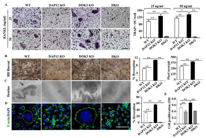
(A) BMMs from WT, DAP12 KO, DOK3 KO and DAP12/DOK3 DKO were cultured in the presence of 20 ng/ml M-CSF and treated with RANKL at concentration of 25 ng/ml and 50 ng/ml. Cells were fixed and stained for TRAP. Representative images were captured by light microscopy and TRAP-positive stained cells (≥ 3 nuclei) were quantified as osteoclasts. (B-D) BMMs were plated and differentiated with M-CSF (20 ng/ml) and RANKL (50 ng/ml). (B) BD Biocoat resorption pits at 7 days. Representative images of resorption pits (white areas) were captured (original magnification, 200×). Percent resorption (%) and pixel area per pit were quantified (n = 100 pits per group). (C) Representative images of dentin resorption pits and percent resorption (%) quantified. (D) Osteoclasts were fixed, permeabilized and stained with Alexa Fluor® 488 Phalloidin to visualize actin rings. (E) MTT assay of proliferation of BMMs from WT, DAP12 KO, DOK3 KO and DKO mice. BMMs were seeded and cultured with M-CSF (20 ng/ml) for 24 hours. Statistical values were calculated using a Student's t-test unless otherwise indicated Scale bar: 100 μm (A and D). Experiments were repeated three times. *p < 0.05, **p < 0.01. Error bars indicate the mean ± SD of triplicates.