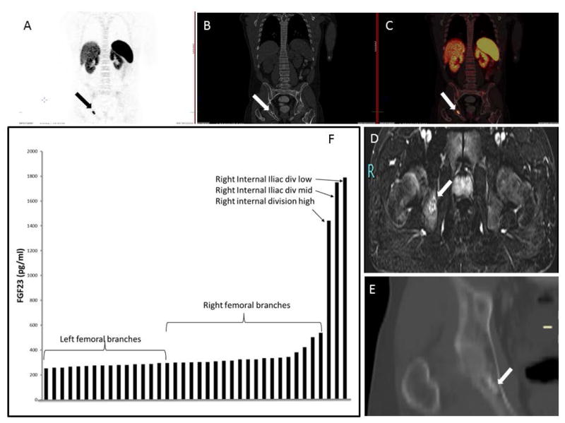
Figure 5.
Localization studies subject 3: Panels A, B, C: Ga68 DOTATATE isotopic uptake in right ischium corresponding to the right ischial mass. Panel D: MRI of the pelvis confirmed the ischial location. Panel E. CT scan coronal view of pelvis confirmed the lesion location in right ischium budding the acetabulum. Panel F. Selective venous sampling for intact FGF23. There was a significant step-up in FGF23 concentration as the suspected tumor venous drainage was approached anatomically.

