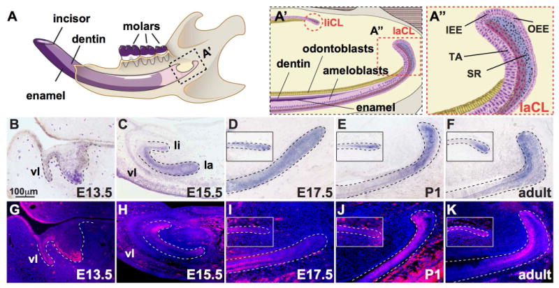
Figure 1. Expression of Isl1 during development of the mouse incisor.

(A) Illustration of the mouse hemi-mandible showing the incisor and molars, as well as the mineralized dentin and enamel comprising the incisor. (A′) The proximal region of the incisor denoting the labial and lingual cervical loop (laCL and liCL, respectively, highlighted by dashed, red lines). (A″) Magnified view of the laCL showing the inner enamel epithelium (IEE), outer enamel epithelium (OEE), stellate reticulum (SR), and transit-amplifying cells (T-A). (B-F) In situ hybridization staining for Isl1 at embryonic day 13.5 (E13.5), E15.5, E17.5, post-natal day 1 (P1), and 6-week old (adult) showed Isl1 expression throughout mouse incisor development including the vestibular lamina (vl), and the lingual (li) and labial (la) aspects. In adults, Isl1 expression was predominant in the laCL and liCL. (G-K) Immunofluorescence staining for ISL1 during mouse incisor development showed similar expression profiles to Isl1 expression (B-F) with the exception of E15.5, where protein expression was increased on the lingual side.
