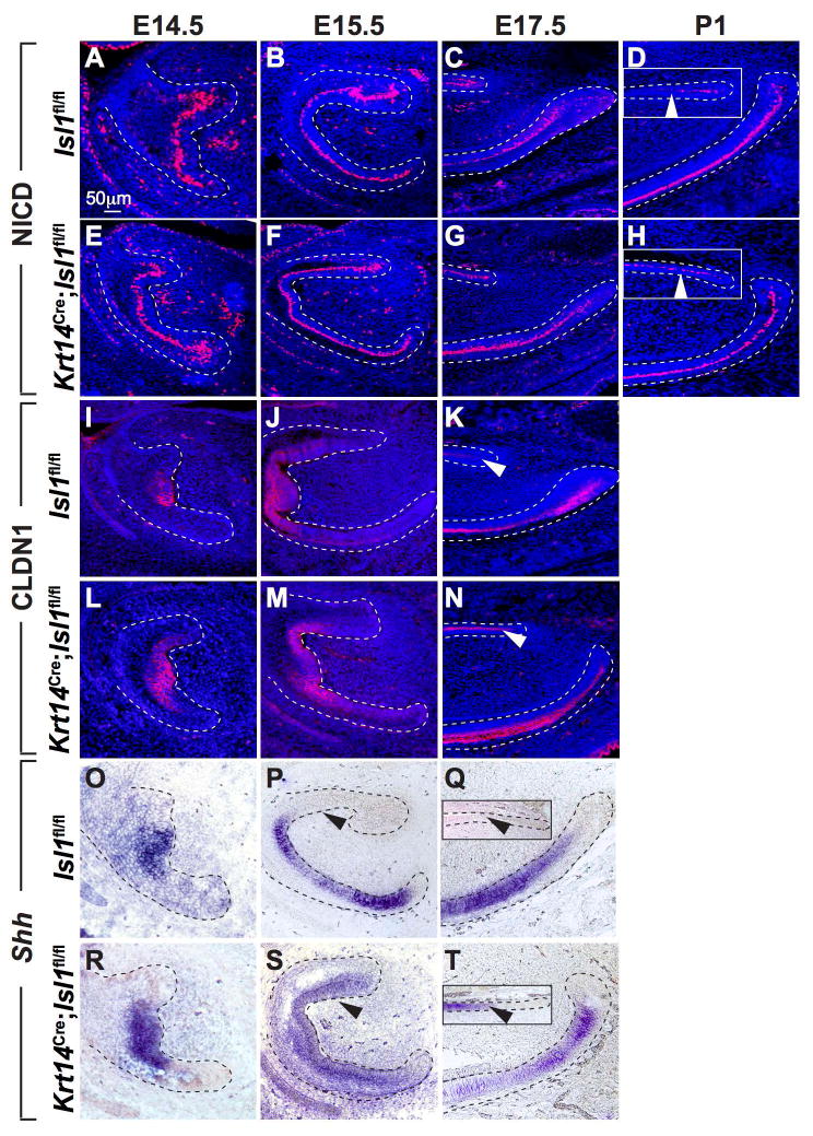
Figure 7. Expression of NICD, CLDN1 and Shh in the CLs during development.

(A-H) Immunofluorescence staining for NICD showed similar patterns of expression in control and mutant mandibular incisors until E17.5 (C,G). At P1, NICD staining decreased in control liCL but remained in the mutant liCL (D,H insets; white arrowheads). There appeared to be little difference in NICD expression between control and mutant laCL. (I-N) Immunofluorescence staining for CLDN1 showed similar patterns of expression in the enamel knot area until E15.5 (I,J,L,M). However, similar to NICD staining at P1 (D,H insets), CLDN1 staining remained high in the mutant liCL but decreased in control liCL (K,N; white arrowheads). Again, there appeared to be little difference in CLDN1 expression in control and mutant laCL. (O-T) In situ hybridization analyses showed similar expression patterns in control and mutant mandibular incisors at E14.5 (O,R). At E15.5, Shh expression was evidenced on the lingual aspect of the mutant incisors but not in controls (P,S). At E17.5, Shh expression was present in mutant but not control liCL (Q,T insets; black arrowheads). At E17.5, consistent with observations in adult laCL (Fig. 5 J-L), there appeared to be decreased expression in mutant laCL compared to controls (Q,T).
