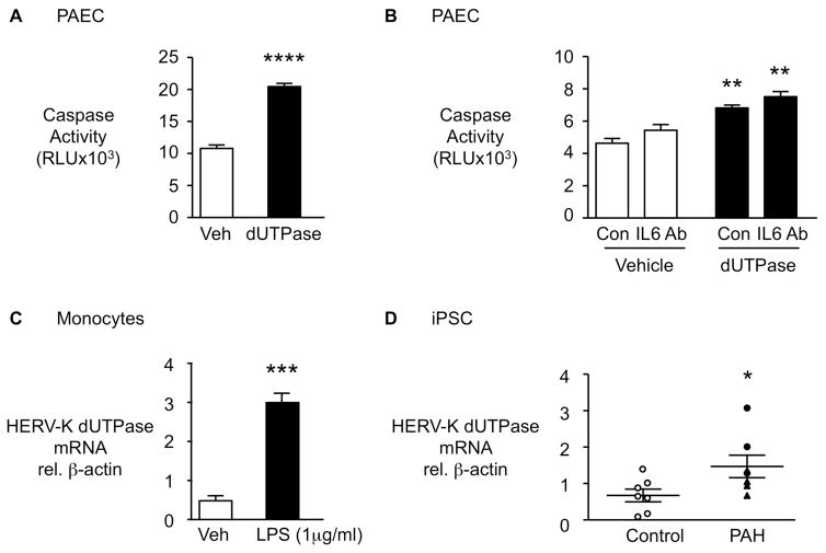
Figure 5. HERV-K dUTPase increases apoptosis in pulmonary arterial endothelial cells (PAEC) in an IL6 independent manner, is induced in monocytes by LPS and is elevated in iPSC from PAH patients vs. controls.
(A) PAEC were treated with 10 μg/mL HERV-K dUTPase and apoptosis was assessed by Caspase-Glo 3/7 assay following overnight serum withdrawal (n=4). (B) PAEC were pre-treated with neutralizing IL6 antibody (IL6 Ab) or isotype control (Con) before HERV-K dUTPase treatment (n=4). (C) HERV-K dUTPase mRNA by qPCR in monocytes stimulated with LPS 1μg/ml (n=3). (D) HERV-K dUTPase mRNA by qPCR in iPSC from PAH patients (n=7) and controls (n=7). Ranges represent mean ± SEM. *P<0.05, ***P<0.001, ****P<0.0001 by Student’s t-test (A, C, D). In (B), **P<0.01, HERV-K dUTPase vs. Vehicle treatment, by one-way ANOVA and post hoc Tukey test. Closed symbols (PAH), open symbols (controls), closed triangles hereditary PAH (HPAH).

