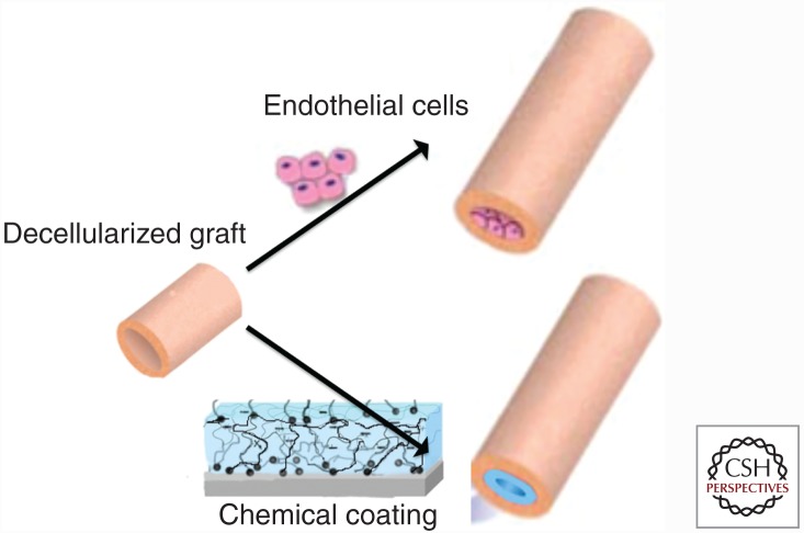
Figure 4.
Schematic representation of the main two approaches for producing an antithrombogenic surface in decellularized vascular grafts. The top drawing represents the biological avenue of seeding endothelial cells (ECs) growing before implantation. The bottom panel represents the chemical strategy, wherein the decellularized surface is “coated,” thereby hiding the collagen from the circulating blood, inhibiting the platelet activation.

