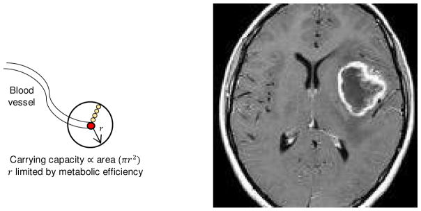
Figure 1.
Local diffusion reaction kinetics around a blood vessel allow microscopic-scale spatial gradients in oxygen, glucose, and H+. Although not spatially explicit, the model equates carrying capacity with the cross-sectional area of a tumor cord growing around a centrally located blood vessel (right panel). At macroscopic scale, the spatial density of blood vessels can vary. The most common pattern is high vascular density in the tumor periphery and low blood flow with tumor necrosis centrally (right panel, ring-enhancing glioblastoma).

